Ketamine and Oxytocin Provide Pain Relief for Arachnoiditis

By Dr. Forest Tennant, PNN Columnist

Nearly every day we receive an email from someone who is having trouble getting enough pain medication to give them a good quality of life.  In this age of opioid restrictions, there is hope. 

In the past, opioids and gamma amino butyric acid (GABA) substitutes such as diazepam (Valium) and gabapentin (Neurontin) have been the mainstays for pain control of adhesive arachnoiditis (AA). Today, there are alternatives that can enhance your current program to give you better pain control.

Low dose naltrexone is the initial pain reliever recommended to newly diagnosed AA cases. There are two other potent pain relievers that can be used with both naltrexone and opioids to achieve better pain relief: ketamine and oxytocin. Either agent is a good opioid substitute.

Ketamine provides pain relief primarily by suppressing a nerve receptor called N-methyl-d-aspartate. It can be taken by several non-oral routes of administration: nasal, injectable, sublingual or troche (dissolvable tablet).

Oxytocin (not to be confused with oxycodone or OxyContin) is a hormone that is a natural pain reliever. It surges in a woman’s body at the time of delivery to provide pain relief. It acts by activating the endorphin (opioid) receptors and by blocking nerve impulses between the brain and spinal cord.

Every community now has one or more pharmacies that will compound or “make” formulations of ketamine or oxytocin. We favor under-the-tongue (sublingual) or buccal (cheek) formulations.

Ketamine and/or oxytocin can be taken between opioid dosages or within 5 to 10 minutes before or after an opioid dosage to make the opioid stronger and last longer.

Ketamine and oxytocin can be used separately or used as combination therapy. Starting dosages of ketamine are 10-15 mg and oxytocin 10-20 units, which are administered within 10 minutes of each other. Dosages can later be raised above the starting dose.

We find the combination of ketamine and oxytocin to provide equal or better pain relief that most prescription opioids. Neither ketamine nor oxytocin are opioids, so there is no bias or resistance to their use. Also, overdoses are essentially not known to occur with regular dosages.

Forest Tennant, MD, DrPH, is retired from clinical practice but continues his research on the treatment of intractable pain and arachnoiditis. This column is adapted from a bulletin recently issued by the Arachnoiditis Research and Education Project. Readers interested in subscribing to the bulletins should click here.

The Tennant Foundation gives financial support to Pain News Network and sponsors PNN’s Patient Resources section.   

Electromedical Treatments for Arachnoiditis

By Dr. Forest Tennant, PNN Columnist  

Adhesive Arachnoiditis (AA) is an inflammatory, nerve root entrapment disease in which cauda equina nerve roots are glued by adhesions to the arachnoid-dural covering of the spinal canal. An inflamed tumor-like mass is formed inside the spinal canal that blocks spinal fluid flow, allows seepage of fluid into tissue outside the spinal canal and shuts off electrical impulses that activate the legs, feet, bladder, intestine and sex organs. Autoimmunity is produced and/or magnified by AA. 

We highly recommend a three-component protocol for AA to reduce inflammation and autoimmunity, regenerate damaged tissue and to provide pain control. Recent advances in electromedical therapies can help achieve these three goals. 

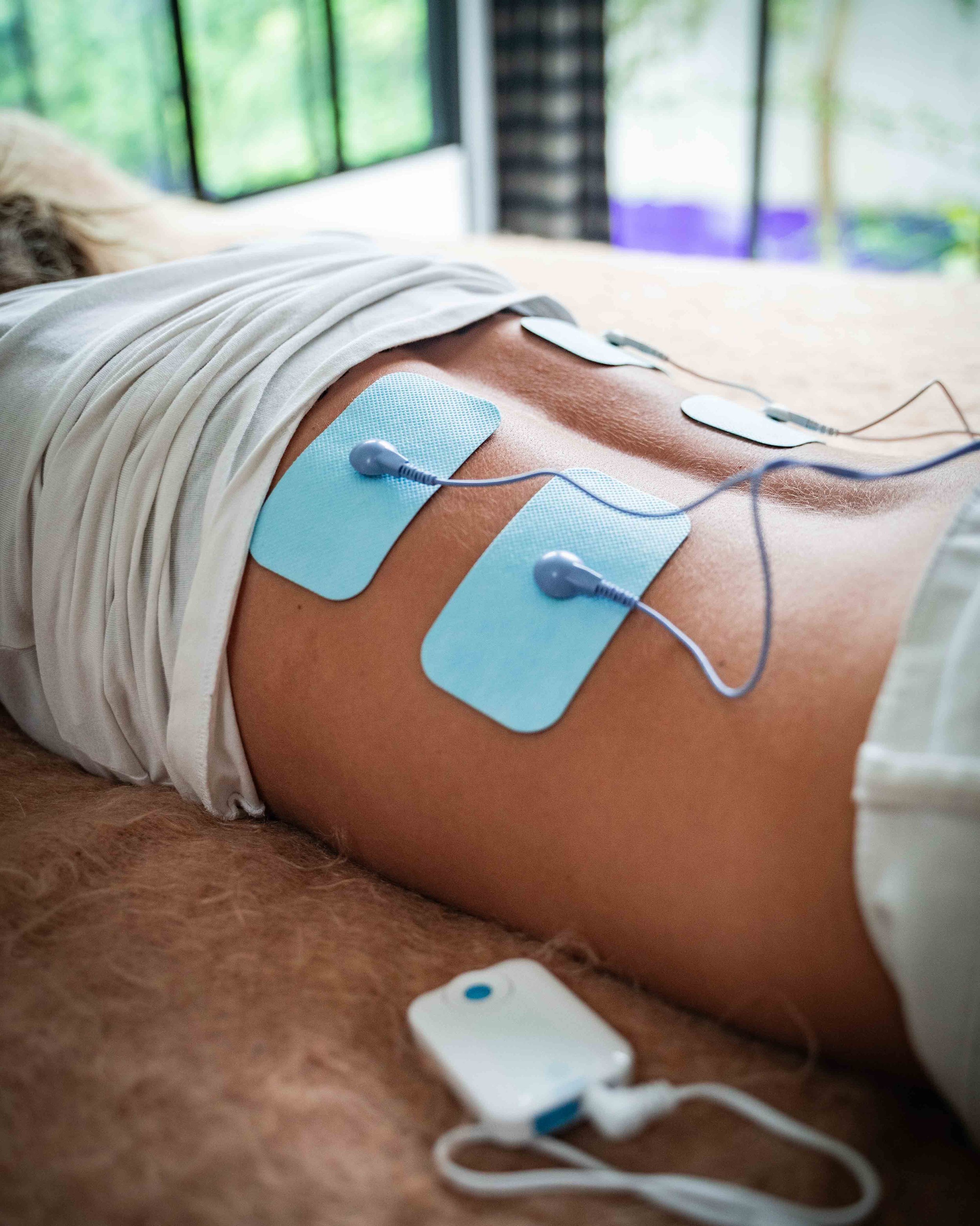
There are two basic types of electromedical devices available for AA treatment: electric current therapy (EC) and electromagnetic therapy (EM). 

Electric Current Therapy 

Almost everyone is familiar with “TENS” units, which stands for “transcutaneous electrical nerve stimulation.” These devices were the first electromedical therapies to relieve pain and promote healing.

TENS units deliver a single electric current into tissues to produce an anesthetic, pain relieving effect.  

Today, more advanced EC devices administer micro-currents and/or a combination of multiple currents with different frequencies. 

Electromagnetic Therapy 

There is a form of energy that is half electricity and half magnetism, which can be divided into wave lengths. The very shortest wave of electromagnetic energy is “atomic” and the longest is “radio.” The shortest wave used in medicine is “laser.” Other electromagnetic energy waves used for medical purposes include infrared, light and microwave. 

EC and EM devices, when placed over the lower back, deliver electric current or electromagnetic energy to the lumbar-sacral spinal canal and the spine’s surrounding tissue.

Modern devices use intermittent pulsation of electric currents or electromagnetic energy to penetrate the skin and subcutaneous tissue to reach the AA site, which is usually about 2-3 inches below the skin.  

Some devices use the label PEMF, which stands for “pulsed electromagnetic frequency.” We believe that the newer EC and EM devices can deliver electric currents or electromagnetic energy that, when pulsed, penetrate deep enough to reach the AA disease site. 

Although not totally curative, these devices usually bring about pain reduction in the 20 to 30% range. Within an individual’s financial capability, we recommend that an EC and/or EM device be used 2 to 3 times a week (not daily). EC and EM therapy are not substitutes for a medical protocol. 

EM and EC devices often produce some initial healing, but later seem to stop working. In this situation the device may have done its maximal healing. The devices can still be used periodically to prevent relapses and treat flares. 

Forest Tennant, MD, DrPH, is retired from clinical practice but continues his research on the treatment of intractable pain and arachnoiditis. This column is adapted from a bulletin recently issued by the Arachnoiditis Research and Education Project. Readers interested in subscribing to the bulletins should click here.

Dr. Tennant’s new book, "Clinical Diagnosis and Treatment of Adhesive Arachnoiditis” is available on Amazon. 

The Tennant Foundation gives financial support to Pain News Network and sponsors PNN’s Patient Resources section.   

Popular Exercises for Persons with Arachnoiditis

By Dr. Forest Tennant, PNN Columnist 

Adhesive Arachnoiditis (AA) is an inflammatory, nerve root entrapment disease in which cauda equina nerve roots are glued by adhesions to the arachnoid-dural covering of the spinal canal. An inflamed tumor-like mass is formed inside the spinal canal that blocks spinal fluid flow, allows seepage of fluid into tissue outside the spinal canal, and shuts off electrical impulses that activate the legs, feet, bladder, intestine and sex organs.  

Some specific exercises help neutralize the deleterious effects of AA and promote regeneration of damaged tissue.  We surveyed 40 persons with MRI-documented AA to determine which exercises they found most beneficial.

The top five are listed here in descending order of popularity. 

  1. Water Soaking: It is no surprise this is No.1. Water soaking pulls out toxins and excess electricity and relaxes muscles. All types of water soaking are good: pool, jacuzzi, shower, tub, hot/wet towel. Epsom Salts in water mimic the mineral baths used therapeutically by ancient peoples. 

  2. Massage: Kneading of back muscles causes any seepage of spinal fluid to mobilize and causes spinal fluid to keep moving around the AA blockage in the spinal canal. 

  3. Walking: Nerve roots that activate the legs and feet can become so inflamed and entrapped that one can’t walk. Short daily walks are essential to prevent the development of paralysis and weakness. 

  4. Arm & Leg Stretching: Entrapped nerve roots in the AA mass decrease the normal leg, arm, and foot fidgets and movements that occur every few minutes even while sleeping. Arm and leg stretching will keep the lower back muscles from contracting or shrinking which, over time, will increase back pain. 

  5. Deep Breathing: Deep breathing and short breath-holds bring oxygen to the spinal canal to promote healing. It will also help keep spinal fluid moving. Deep breathing is best done while standing but it can be done while sitting and watching TV, driving, or eating. 

Other exercises compliment the AA medical treatment protocol. Besides those listed here, we also advocate light weightlifting, rocking, bicycling, and trampoline walking. 

Credit: Lynn Ashcraft did the data analysis of this survey. 

Forest Tennant, MD, DrPH, is retired from clinical practice but continues his research on the treatment of intractable pain and arachnoiditis. This column is adapted from a bulletin recently issued by the Arachnoiditis Research and Education Project. Readers interested in subscribing to the bulletins should click here.

Dr. Tennant’s new book, "Clinical Diagnosis and Treatment of Adhesive Arachnoiditis” is available on Amazon. 

The Tennant Foundation gives financial support to Pain News Network and sponsors PNN’s Patient Resources section.   

Emergency Treatment of Arachnoiditis After Spinal Injection

By Dr. Forest Tennant, PNN Columnist

In rare cases, symptoms suggesting adhesive arachnoiditis (AA) may occur after a spinal tap or epidural injection (therapeutic or obstetrical). These early symptoms may include localized lumbar pain, headaches, burning sensations, dizziness, leg weakness and bladder dysfunction. Spinal fluid leaks or blood in the spinal canal are often suspected in these cases.  

If symptoms indicate the possibility that AA may be developing, we recommend emergency treatment to hopefully prevent the spinal nerve inflammation from spreading and becoming chronic. 

A problem that we have routinely discovered is that medical practitioners commonly have the false belief that they can see signs of AA on an MRI when symptoms begin or within a few hours or days after a spinal tap or epidural injection. But AA typically does not show on an MRI for at least four to six weeks. Consequently, early emergency treatment must be based on patient history and symptoms, rather than on MRI findings. 

At the First International Congress on Arachnoiditis and Tarlov Cysts in 2010, physicians Donna Holder and Antonio Aldrete recommended that methylprednisolone 500 mg be given intravenously every day for five days as an emergency treatment for AA.  

Since that time, a variety of intravenous methylprednisolone attempts with different dosages and frequency have been used by physicians as emergency treatment to prevent AA. Dr. Aldrete opined that intravenous methylprednisolone is only effective in preventing AA if given within 60 days after the spinal tap or epidural. 

We have used the following alternative treatments to intravenous methylprednisolone: 

  1. Medrol (methylprednisolone) six-day oral dose pack

  2. Ketorolac 30 to 60 mg injection for three consecutive days

  3. Medroxyprogesterone 10 mg given twice a day for six days

In some, but not all cases, AA symptoms will abate during the week that either intravenous methylprednisolone or the alternative treatments are administered. In most cases, however, symptoms reduce but don’t totally abate. The reason for this is unclear, but a reasonable assumption is that spinal canal inflammation may not be totally reversed once symptoms begin. 

If pain and other symptoms don’t totally abate, we recommend that the patient begin a three-component medical protocol for AA described in this bulletin, which includes nutritional, physiologic and pharmacologic elements. Patients should remain on these medical treatments until and if their pain and other symptoms resolve. 

It is unclear why only a small percentage of persons who have spinal taps or epidural injections develop AA. It is also unknown why symptoms that begin after these procedures usually don’t abate. 

A New Handbook for Practitioners

Our new book, "Clinical Diagnosis and Treatment of Adhesive Arachnoiditis” is now available on Amazon. 

This handbook for medical practitioners has been written for one simple reason.  AA is no longer a rare disease.  We conservatively estimate that there are at least 1.75 to 2.75 million adults in the U.S. who have AA.  

In the past, the cause of their back pain was often listed as unknown or inappropriately labeled as failed back syndrome, degenerative spine or simply low back pain. 

It is our fervent hope that this book will help medical practitioners and their patients diagnose and treat this most debilitating disease.

Forest Tennant, MD, DrPH, is retired from clinical practice but continues his research on the treatment of intractable pain and arachnoiditis. This column is adapted from a bulletin recently issued by the Arachnoiditis Research and Education Project. Readers interested in subscribing to the bulletins should click here.

The Tennant Foundation gives financial support to Pain News Network and sponsors PNN’s Patient Resources section.   

How the Epstein Barr Virus Causes Arachnoiditis

By Dr. Forest Tennant, PNN Columnist

The Epstein-Barr Virus (EBV) has long been known to cause autoimmune complications. For example, I described infectious mononucleosis in kidneys (glomerulonephritis) associated with EBV back in 1969, some 53 years ago.

Recently EBV has been shown to cause multiple sclerosis (MS), as well as certain cancers. Our studies have also revealed that many, if not the majority, of persons with adhesive arachnoiditis (AA) have extremely high EBV antibody levels. We now believe that EBV can cause AA as well as MS. 

Anything that boosts or helps a disorder to develop, is called a “co-factor.” In the case of EBV and AA, the “co-factors” or “co-partners” may include Ehlers-Danlos Syndromes, autoimmune diseases such as psoriasis, Lyme disease, and some other viruses like cytomegalus, coxsackie and covid.

‘Collagen Eaters’

Although almost everyone gets infected with EBV during their lifetime, some persons develop a large number of EBV antigens that literally attack and “eat away” or otherwise cause collagen to deteriorate and weaken. If the body creates large numbers of antigens, large numbers of antibodies are simultaneously made to hopefully counter them.

EBV collagen eating antigens like to attack tissues around the spine that have a lot of collagen. This includes intervertebral discs, cauda equina nerve roots, and the arachnoid-dural covering of the spinal canal. If collagen is weak or absent in spinal tissues, discs may slip, inflammation may form, and fluid leaks and cysts may develop.

Get Tested for EBV

All persons with confirmed or suspected AA should be tested for EBV antibodies.

Laboratories today do three types of tests for EBV. One is to tell if you currently have an active infection and the other two measure antibody levels to determine if you have had EBV in the past. If your antibodies are considerably above normal range, it suggests you have collagen eating antigens that put you at risk of developing AA.

Forest Tennant, MD, DrPH, is retired from clinical practice but continues his research on the treatment of intractable pain and arachnoiditis. This column is adapted from a bulletin recently issued by the Arachnoiditis Research and Education Project. Readers interested in subscribing to the bulletins should click here.

The Tennant Foundation gives financial support to Pain News Network and sponsors PNN’s Patient Resources section.  

How to Improve Pain Control with Adhesive Arachnoiditis

By Dr. Forest Tennant, PNN Columnist

Some persons with Adhesive Arachnoiditis (AA) feel that more pain relief drugs, particularly opioids, will solve their problem. The fact is that we regularly review cases in which persons with AA are taking 2 or 3 opioids or even have an implanted intraspinal canal pump that contains fentanyl or Dilaudid, but they still don’t get enough pain relief to get out of bed and function.

AA is an intraspinal canal inflammatory disorder in which cauda equina nerve roots are glued by adhesions to the inside of the spinal canal covering. This definition tells it all. AA is fundamentally intraspinal canal inflammation, so unless the initial inflammation is suppressed or extinguished, it will likely continue to spread and cause more pain.

In essentially every case of failing pain control, we find that the person is doing little or nothing to suppress intraspinal canal inflammation and repair damage to their nerve roots and spinal canal covering.

First Steps in Pain Control

Our research is clear. A person with MRI-documented AA can’t expect adequate pain relief unless they have a definite, daily routine to simultaneously suppress intraspinal canal inflammation and repair tissue damage to cauda equina nerve roots and the spinal canal covering.

Adequate pain control to have a good quality of life can be difficult to achieve. The first step is to obtain a list of drugs, botanicals, hormones, nutrients and physical measures that are popular in the AA community and that either suppress inflammatory or restore damaged tissue. Share your list with your family and medical practitioner. You may have to try multiple agents to develop a program that gives you better pain control.

Persons who have AA and poor pain control also need a blood test for inflammatory markers, glucose, and the hormones cortisol, pregnenolone, DHEA and testosterone.

If your pain is constant, review our Intractable Pain Syndrome website that is totally dedicated to relief from constant pain.

How to Diagnose AA

I’m pleased to announce the release of a new handbook that takes the mystery out of diagnosing AA with contrast MRI imaging once and for all. I have read hundreds of contrast MRI’s during my years in medical practice, and have found that the earlier a diagnosis is made and treatment is started, the better the prognosis is for the patient.

Unfortunately, many health care practitioners don’t know the telltale signs of AA when it appears in an MRI. As a result, AA is often misdiagnosed as “Failed Back Syndrome” or “Low Back Pain.”

“Handbook to Recognize Adhesive Arachnoiditis” is an essential read for all practitioners who are interested in treating patients with spine disorders and patients who suspect they may have AA. It’s presented in a clear and easy to read format as a “how to” guide for reading contrast MRI’s for the diagnosis of AA.

The book is filled with clearly diagrammed MRI images of documented cases of AA and should help practitioners diagnose AA and learn the difference between AA and other spine disorders with similar symptoms.

This book will also help those patients who suspect they may have AA receive a quick and proper diagnosis, thus preventing delays in effective treatment of this devastating spinal cord disease. AA is no longer rare. It is in every community, and health care practitioners can now learn how to diagnose and treat it. There is hope and help!

Forest Tennant, MD, DrPH, is retired from clinical practice but continues his research on the treatment of intractable pain and arachnoiditis. This column is adapted from a bulletin recently issued by the Arachnoiditis Research and Education Project. Readers interested in subscribing to the bulletins should send an email to tennantfoundation92@gmail.com.

The Tennant Foundation gives financial support to Pain News Network and sponsors PNN’s Patient Resources section.  

Is Your Spinal Pain Inflammatory or Neuropathic?

By Forest Tennant, PNN Columnist

Every person with Adhesive Arachnoiditis (AA) or other spinal canal disorder needs to determine if their pain is primarily inflammatory, neuropathic or both. Why? The treatments are different.

AA is fundamentally an inflammatory disease that involves two different intraspinal canal tissues: the cauda equina nerve roots and the arachnoid-dural covering of the spinal canal. The inflammation causes damage to the nerve roots, so electricity either can’t pass or it doesn’t pass in a smooth, natural flow.

Nerve damage that blocks or alters electricity conduction is called “neuropathic” pain. AA usually has both inflammatory and neuropathic pain, but the inflammation may resolve and leave behind damaged nerve roots and neuropathic pain.

The inflammatory and neuropathic pain of AA may also develop into Intractable Pain Syndrome, which is constant, incurable pain with cardiovascular, endocrine (hormonal) and autoimmune complications.

Persons with AA usually need to treat both kinds of pain – inflammatory and neuropathic --   but one type may be predominant. A blood test for inflammatory markers is helpful, but not totally diagnostic.

If your pain improves with a trial of ketorolac (1 or 2 injections) or a corticosteroid (Medrol Dose Pak or dexamethasone), you have active inflammation that must be treated. We also recommend botanical anti-inflammatory agents, such as curcumin/turmeric, Andrographis and serrapeptase.

Prescription medications for neuropathic pain include gabapentin (Neurontin), diazepam, carisoprodol, topiramate, Lyrica and Cymbalta.

Every person with AA of the cervical and/or lumbar spines should experiment with topical medications, such as the Salonpas patch, lidocaine gel or patch, Voltaren gel and diclofenac (prescription needed).

Topical medication that is applied and massaged into the skin may dissolve through the tissues to the inflamed or damaged area. On average, you can expect 10 to 25% additional pain relief, plus the potential to permanently reduce your pain. Sometimes topical  medication will relieve painful areas that other drugs taken orally or by injection cannot reach.

Forest Tennant is retired from clinical practice but continues his research on the treatment of intractable pain and arachnoiditis. This column is adapted from bulletins recently issued by the Arachnoiditis Research and Education Project. Readers interested in subscribing to the bulletins should send an email to tennantfoundation92@gmail.com.

The Tennant Foundation gives financial support to Pain News Network and sponsors PNN’s Patient Resources section.  

Great Progress Being Made in Treating Arachnoiditis

By Dr. Forest Tennant, PNN Columnist

About 5 years ago, most medical practitioners had either never heard of Adhesive Arachnoiditis (AA) or thought it was a spider bite. Today, almost all practitioners in the modern world have heard of AA. Many now understand it and some even treat it. A few are trying some innovative new approaches.

AA is a chronic inflammation that starts inside the spinal canal that can lead to severe suffering, neurologic impairments and a shortened lifespan. Once inflammation starts, it apparently never, or rarely, goes totally away.  

Treatment and prevention in recent years have greatly reduced the occurrence of some serious neurologic impairments and autoimmune complications of AA. The most obvious decrease in new cases reviewed by the Tennant Foundation are those of upper and lower extremity paraparesis (partial paralysis) and total paralysis, which are rapidly disappearing.

Urinary and bladder impairments that require catheterization are also hardly seen. And the autoimmune manifestations of arthritis, thyroid deficiency and carpal tunnel are disappearing.

Why the improvement? Awareness, thanks to patients, social media and advocates who have educated the medical profession about AA. Fewer epidurals, early treatment and emergency measures have all helped. The development of protocols for prevention, emergency intervention and on-going treatment have been essential.

Major Remaining Problems

Persons with AA are still having difficulty, in some communities, finding medical practitioners who are comfortable and willing to treat AA. The major complication is the development of constant pain and the intractable pain syndrome.

The key to preventing AA and stopping its progression is early treatment. Our research has clearly shown that AA is almost always preceded by one of 3 intraspinal canal inflammatory conditions:

  1. Protruding, degenerated intravertebral discs.

  2. Cauda equina inflammation.

  3. Arachnoid inflammation (i.e. plain arachnoiditis) due to collagen disorders or needle injury.

Some intraspinal canal inflammatory disorders always precede AA. These disorders should be aggressively treated to prevent AA.

Select Corticosteroids Essential for AA

We believe all persons with typical AA symptoms and documentation of the disease on an MRI must take one of two corticosteroids (CS): methylprednisolone or dexamethasone for the spinal canal inflammation and pain of AA.

Currently there is no other medication agent that consistently and predictably suppresses intraspinal canal inflammation and reduces pain. Do not expect to halt progression or have much recovery if you do not consistently take a CS.

Dexamethasone and methylprednisolone are the preferred CS’s because they cross the blood brain barrier, enter spinal fluid and act on glial cells. Prednisone and hydrocortisone are not as consistently effective as dexamethasone and methylprednisolone, which should be taken in low doses.

  1. Maintenance-low dose of dexamethasone (.5 to .75mg) or methylprednisolone (Medrol) 2 to 4 mg on 2 to 5 days a week. Skip days between dosages. An alternative is a weekly or bi-monthly injection of methylprednisolone or dexamethasone. Injections are usually the answer to corticoid sensitivity or gastric upset.

  2. For flares, a 6-Day Medrol Dose Pak or an injection of methylprednisolone or dexamethasone, preferably mixed with a standard dose of injectable ketorolac.

The fear of corticosteroids comes from daily use of high doses, not from low, intermittent dosages. Some persons with severe asthma and rheumatoid arthritis must take a corticosteroid for years and don’t experience serious side effects.

Forest Tennant, MD, MPH, DrPH, is retired from clinical practice but continues his groundbreaking research on the treatment of intractable pain and arachnoiditis. This column is adapted from bulletins recently issued by the Arachnoiditis Research and Education Project of the Tennant Foundation. Readers interested in subscribing to Dr. Tennant’s bulletins should send an email to tennantfoundation92@gmail.com.

Dr. Tennant and the Tennant Foundation have given financial support to Pain News Network and are currently sponsoring PNN’s Patient Resources section.  


The Other Side of Chronic Pain

By Donna Corley, Guest Columnist

As I contemplated how to begin this article on a subject that has troubled me for some time, I continue to hear Paul Harvey’s famous tag line, “And now you know the rest of the story.”

I’ve noticed for a while that when we share our stories about life with chronic pain, we often focus on the physical struggles and lack of proper treatment, but leave out the emotional side. We rarely tell anyone how vulnerable and afraid we feel, especially our families and doctors, for fear of them thinking we are faking it or even a little crazy.

I’ve hidden that side of myself very well -- as have many of you -- and for good reason. But in doing so, I’ve only told half of my story and only shared half of who I am.

It is very hard to show vulnerability to anyone because we are judged by so many people. No one wants to be ridiculed for how they feel.

We should not be made to feel this way. None of us asked for pain conditions and diseases. Yet, we are shunned and doubted, and made to feel like we should not talk about the emotional side of our journey or the fear of “what comes next.”

That is the secret we hide from the world.

DONNA CORLEY

DONNA CORLEY

My story is a little unique in the sense of how long it has been. My journey started my senior year of high school, when I broke my back in a bad car accident. I was unaware that one accident could manifest into conditions called Adhesive Arachnoiditis (AA) and Tarlov Cyst disease. It took 23 years before I was properly diagnosed with AA. I still have moments when I think, “If only I was diagnosed sooner.”

I think there’s a big misconception about patients who suffer with chronic pain conditions, especially those with no cure. The public doesn’t realize that there is more to us than our pain. But if we don’t talk about our emotional torment, how could they know?

Living in Fear

Being a mother brings worry naturally. When you add massive pain with weird symptoms that make you feel like an alien has taken over your body, well that’s fear. Full-fledged, terrifying “I’m dying” fear. Fear brings anxiety, which brings doubt, and this can bring on depression. And it all started with pain no doctor could explain or diagnose, let alone treat!

The medical community is still being taught that Tarlov Cyst isn’t worth looking for in an MRI because they believe the disease doesn’t cause pain or other symptoms. That’s not true in my case at all. I went years before Tarlov Cyst was diagnosed and only because part of my pain couldn’t be explained. Even then the physicians refused to admit the Tarlov Cyst was causing any symptoms.

I can remember when my children were little having anxiety so severe that I would hyperventilate. Fear gripped me worse than the pain. Fear of dying and leaving my children. Fear that I had something terminal. No doctor wanted to dig deeper to find out what was wrong with me. My pain would be so severe at times it felt as if someone was ripping my spinal cord out, yet I kept this to myself.

I never told anyone the extent of my pain for fear of people thinking I was exaggerating, or worse, that it was all in my head. I quit driving for several years because I was scared my pain would become so severe I wouldn’t be able to make it home.  

Looking back, I see how pain ruled my life to the extent that it brought on major anxiety. I missed out on so many things with my children, their school functions, church, ball games, etc., all because of pain, fear and anxiety.

You learn how to hide your pain. Even if it means sneaking off to the bathroom to cry for a few minutes because you are hurting so much. You become a pro at clenching your jaw and smiling through the pain. Constant pain keeps us in a “fight or flight” state that can be very hard to manage alone.

I’ve seen doctor after doctor more times than I can count. They would all ask me, “Why are you here?” I finally stopped going. I stopped asking my primary care physician to refer me. What was the point? Physicians used to look outside the box to find answers, but it seems that “patient centered care” is gone. I had a doctor tell me a couple years ago that he couldn’t help me because I had too many health problems!

Doctors can’t always fix us, but their words and actions have lasting consequences, and some are just resoundingly bad.  Many patients have developed severe emotional trauma from being bullied by physicians who talk down to them like they were stupid and insignificant.

There is another important subject that everyone shies away from, even in patient support groups, and that’s sex. I can understand it being a sensitive subject, but it affects more patients than people realize. Whether it’s erectile dysfunction, the inability to have sex because of pain, or the inability to have an orgasm, these conditions can tear marriages and relationships apart. But the subject is not often discussed with physicians or even family members.

I had to give up a job and career I loved due to chronic pain and had to find a new purpose. That was a very hard part of my life. First, it was hard accepting that I could no longer work, and then it was hard accepting that I would be in pain for the rest of my life.

I thought it was a death sentence when I was first diagnosed with AA. Then I realized, “Hey, I’ve had it for 23 years and I’m not dead yet.” Being a quitter isn’t in my DNA, and I love my family too much to quit. I did a lot of praying, asking God, “Why me?” It was like he said to me, “I need you here.”

That’s truly when I began to have a passion, a drive to fight for awareness, to help other patients get a timely diagnosis and find new doctors.  Praise God, we have quite a few great ones, but we need more.

No one should be made to feel like they are crazy, ignorant, insignificant or lying because they are in pain. I’m still here trying to love the life God has blessed me with. Yes, I see my life as a blessing. If I had not been diagnosed with these diseases, I wouldn’t know how to empathize with others in pain.

We as patients should be able to discuss our fears and concerns with our physicians and families without fear of negative consequences. It is past time people quit condemning those of us who suffer in pain because of the way we feel.

Donna Corley is the director of the Arachnoiditis Society for Awareness and Prevention (ASAP) and creator of the Tarlov Cyst Society of America. She lives in Mississippi with her family.

Pain News Network invites other readers to share their stories with us. Send them to editor@painnewsnetwork.org.

Patients with Arachnoiditis and Ehlers-Danlos Need Adrenaline for Pain Control

By Dr. Forest Tennant, PNN Columnist

In this era of opioid controversy and tragedy due to forced opioid reduction, the scientific information on adrenaline-type agents can help control constant, intractable pain and help reduce opioid use.

Overlooked in the opioid controversy is the key point that an adequate supply of adrenaline-related neurotransmitters – such as dopamine and norepinephrine – are necessary in the brain and spinal cord for pain relief. Unfortunately, constant intractable pain depletes the natural supply of endorphin, dopamine, adrenaline and noradrenalin, and their levels must be replaced to adequately control pain.  

Persons with a severe, intractable pain condition like Adhesive Arachnoiditis (AA) and/or a genetic connective tissue disorder like Ehlers-Danlos syndrome (EDS) will need an adrenaline agent, also called a stimulant, for pain control, maximal function, keeping opioid dosages stable, and preventing sedation and overdose. 

Adrenaline agents have long been known to boost or potentiate opioids, enhance pain relief, and allow less opioid to be used. A study by this author found that the simultaneous use of a stimulant and clonidine lowered opioid dosage by 30 to 50 percent. 

The use of a stimulant in this manner is not new. In the 1920’s, physicians at the Royal Brompton Hospital in London found that a stimulant was a necessary ingredient in the famous “Brompton Cocktail” for relief of severe pain. Today, modern pain relief agents often add caffeine as a stimulant to make the codeine and oxycodone more potent.

Most observers believe that an adrenaline agent given to an intractable pain patient will automatically raise blood pressure and pulse rate. This is generally a myth, because the person with intractable pain often depletes their reserve of dopamine, noradrenalin and adrenaline.  

The use of an adrenaline agent will serve to replace these depleted neurotransmitters and will not generally cause blood pressure and pulse rate to rise. Periodic monitoring is, however, recommended to be continued.  

Multiple Benefits  

A person with intractable pain due to AA, EDS, Reflex Sympathetic Dystrophy (RSD), or another severe and tragic condition will usually have the following conditions – all of which will benefit by an adrenaline agent:

  • Weight Gain

  • Attention Deficit Disorder (ADHD)

  • Depression

  • Sedation

  • Fatigue

  • Memory Loss

Descending Pain

Recent research has learned that constant, intractable pain establishes a “biologic battery” in the brain and spinal cord. This “battery” sends electric currents down the autonomic (non-spinal cord nerves) nervous system. Symptoms of this descending pain include excess heat, muscle spasms, jerking, tremors, sweating and “all-over” pain.  

In contrast to other forms of pain, descending pain isn’t well controlled by opioids and anti-inflammatory agents. The drugs clonidine and tizanidine are less effective. Only adrenaline agents stop it. Some adrenaline agents for persons with AA and EDS include:

  • Phentermine

  • Adderall

  • Phendimetrazine

  • Methylphenidate

  • Dexedrine

  • Modafinil  

Every person with intractable pain due to AA, EDS, RSD, cancer or other painful disease, should educate themselves on adrenaline agents and discuss them with their medical practitioners in order to either lower their opioid dosage or keep it from escalating.   

Simply stated, a person with intractable pain needs at least a small dose of an adrenaline agent for pain relief and optimal function. 

Forest Tennant, MD, MPH, DrPH, has retired from clinical practice but continues his groundbreaking research on the treatment of intractable pain and arachnoiditis. This column is adapted from a bulletin recently issued by the Arachnoiditis Research and Education Project of the Tennant Foundation, and is republished with permission. Correspondence should be sent to veractinc@msn.com.  

Dr. Tennant and the Tennant Foundation have given financial support to Pain News Network and are currently sponsoring PNN’s Patient Resources section.  

Praying for a Miracle

By Stephanie King, Guest Columnist

Up until May of 2010, everything was pretty easy going as far as my health was concerned. I was a happily married 22-year old and mother of a sweet one-year old boy. Then one fateful afternoon all of that changed.

There was a bad storm that day but we had to pay bills. My family and I ended up hydroplaning off the road and crashing into trees.

Luckily, our son was unharmed and my husband, while it was bad, suffered nothing worse than a broken leg.

I had the worst of the injuries and needed surgery. I broke my right foot in three places and my back in five. One of those breaks was a compression burst fracture, which means that my vertebrae basically exploded from pressure. 

Unfortunately, I also developed MRSA, a severe and dangerous bacterial infection. This meant packing the open, oozing wounds every day. Instead of getting better, the infection spread through my back and within a couple of months turned into sepsis. Two more surgeries and six months later, I was finally infection free.

A few years later, my pain was increasing due to degenerative disc disease and arthropathy, so my pain specialist started pushing epidural steroid injections. I was already on fentanyl and MS Contin, so I figured I would give it a shot. This would be the biggest mistake of my life.

I ended up having about ten injections. The last one caused extreme levels of pain and my body felt strange that night. I refused to get any more after that. 

Not long afterwards, I began developing neurological pain that I wasn't used to. Sometimes it felt like warm water running down my legs. Sometimes it was stabbing and biting sensations. Sometimes it felt like my bones were snapping. I didn't make the connection between the epidural and the nerve pain. I assumed it was a pinched nerve, because my back was so messed up. 

STEPHANIE KING

Then in the fall of 2016, my inner calves began to go numb. The numbness rapidly spread up my legs and into my groin. My GP admitted me into the hospital and I had an MRI done.

That day I heard the words I will never forget, the words that changed my life forever: Adhesive Arachnoiditis.

It was explained to me that this is a disease where scar tissue is engulfing spinal nerves, blocking the flow of spinal cord fluid because of inflammation of the arachnoid lining around the nerves.

The more I learned about Arachnoiditis, the more I realized I was doomed to a lifetime of unrelenting pain, a level of pain some doctors have compared to that of bone cancer. I learned of the possibility of paralysis. I learned how there is no cure, just medication to try to manage the disease symptoms and slow down its spread.

I was devastated but kept pushing on, ignoring the pain as much as possible.

Just over a year later, I learned something else. My birth control had failed. I was pregnant with our daughter. My pain specialist immediately dropped me. My obstetrician panicked and tried to get me in with another pain management doctor due to fear of a miscarriage.

I kept hearing how no one would see someone who was pregnant and not already an established patient at their practice. I was referred to a neurologist, but he told my doctor that there was nothing that he could give me that would be safe during pregnancy.

During my pregnancy, the Arachnoiditis symptoms increased tenfold. The burning in my feet and hands became unbearable. I'm one of the "lucky" Arachnoiditis patients who has scar tissue far enough up their spine to effect my arms and hands as well. I began jerking uncontrollably and developed tremors. I spent most of my pregnancy crying and screaming.

We have a beautiful daughter now! She is so sweet and smart! Unfortunately, my symptoms never subsided. I still don't have a pain management doctor. No pain specialist will see me.

I fear I was red flagged for doctor shopping, even though it was just my obstetrician trying to get me treatment. I have finally started seeing a neurologist but until he has reviewed all of my MRIs, no treatment will be given. I won't see him again for another month.

I live in never-ending pain. I rely on family a lot to help care for my children while my husband works. I have come to realize I have no good options at this point. I can continue on in pain and being a total burden to my family, just becoming more burdensome over time.

I could turn to marijuana but it is illegal in my state. My children could be taken away and I could go to prison. I could commit suicide but that would scar my children and further hurt the rest of my family. I am stuck.   

There are times I pray for death. I pray for the body I once had. I pray I could be the mother and wife I once was. I pray for anything to make it stop, even for just a little while. I pray for a miracle.

Stephanie King lives in Alabama.

Pain News Network invites other readers to share their stories with us. Send them to editor@painnewsnetwork.org. 

The information in this column is for informational purposes only and represents the author’s opinions alone. It does not inherently express or reflect the views, opinions and/or positions of Pain News Network.

An Old Disease Returns in Modern Times

By Pat Anson, PNN Editor

Dr. Forest Tennant may no longer be practicing medicine, but he continues his groundbreaking research into the causes and treatment of adhesive arachnoiditis (AA), a chronic, progressive and painful inflammation of spinal nerves.

Once a rare disease linked in the 19th century to infections such as syphilis and tuberculosis, arachnoiditis is re-appearing in surprising numbers and for completely different reasons. Tennant believes millions of Americans may have developed AA, which is often misdiagnosed as “failed back syndrome” – chronic back pain that fails to respond or grows worse after invasive spinal procedures.

“I think it’s now the number one cause of really severe pain in the country. I don’t think there’s any question about that,” says Tennant. “When I closed my clinic, about 70% of the people in there who had to take high dose opioids had an issue with arachnoiditis.”

Tennant and co-author Ingrid Hollis have written a new book -- “Adhesive Arachnoiditis: An Old Disease Re-Emerges in Modern Times” -- to help doctors and patients recognize the symptoms of AA and to offer hope for treating a disease long thought to be incurable.

Why is AA re-emerging?  Tennant says a “perfect storm” began to appear around the year 2000, when longer lifespans, obesity and sedentary lifestyles contributed to more cases. Lack of exercise has long been associated with chronic back pain.

Arthritis, accidents, repetitive stress injuries and even Lyme disease can also cause degenerative spinal conditions that lead to AA.

“But the real factor contributing is probably sedentary lifestyles. Look at the number of people spending 6 to 8 hours a day in front of a computer or TV screen,” says Tennant. “I’m really trying to bring attention to the fact that people need to be aware that this is something that can happen because of our lifestyles.”

As more Americans sought treatment for back pain, millions were given “interventional” procedures such as epidural steroid injections and spinal surgeries – a trend that appears to have increased as opioids fell out of favor. While most interventional procedures do not result in complications, all it takes is a single misplaced needle or scalpel to damage the spinal cord and trigger a lifetime sentence of AA.

“Everybody wants to blame the epidurals and the surgeries for these problems, but they were only doing these things when there were medical indications for them. And if they do the surgeries and the epidurals, a certain percentage of them are going to get arachnoiditis,” Tennant told PNN.

“There is a risk/benefit to these procedures. If you start going in and operating on the spine or you have half a dozen epidural injections, you start increasing your risk for developing adhesive arachnoiditis. One of the points I want to make is that it is the underlining spinal conditions that are causing the problems. The interventions and the surgeries may accelerate it, but the condition was already set.”

For many years, Tennant was one of the few doctors in the U.S. willing to treat AA patients with high doses of opioids. Intractable pain patients from around the country traveled to California to see Tennant – which led to his home and office being raided by the DEA in 2017. No charges were ever filed against Tennant, but the stress of being investigated led to his retirement from clinical practice.

Tennant now consults with other doctors and is developing new protocols for treating AA with hormones and drugs that reduce pain and inflammation while stimulating nerve growth. His book includes a self-screening test for patients and a list of AA symptoms.

Most essential in any AA diagnosis is a good MRI of the spine. Tennant has a library of over 300 MRI’s sent to him by patients and doctors in dozens of countries — a testament to how AA is re-emerging worldwide.

(Dr. Tennant and the Tennant Foundation have given financial support to Pain News Network and are currently sponsoring PNN’s Patient Resources section.)  

16 Key Findings about Arachnoiditis

By Forest Tennant, MD, PNN Columnist

We initiated the “Arachnoiditis Research Project” about 6 months ago. Our first goal was to pull together what we have learned to this point. While we continue to gather new information, this short report is an interim attempt to get our findings into the patient and practitioner communities.  

This report is not intended to be a formal protocol or guideline, but a way to pass on what we have found and determined during the course of our learnings.  Please keep in mind that research is neither static nor absolute.  In the future, newer findings will likely both clarify and expand upon our initial findings as presented in this report.    

Frankly, the response to the Arachnoiditis Research Project has been overwhelming. Each day we receive inquiries from patients and practitioners. Patients want help. Practitioners want to know what to do.

We have now reviewed over 300 MRI’s of Adhesive Arachnoiditis (AA) cases. We have received inquiries from 5 continents and over 17 countries. One thing is clear. The need to research and identify treatment for AA is here.  

The goal of our research is to bring AA treatment to every community worldwide. How? By developing both diagnostic and treatment protocols that can be implemented by any medical practitioner in every community. Here is what we have learned so far:

16 Findings about Adhesive Arachnoiditis  

  1. Treatment efficacy is best achieved by the simultaneous administration of a three component medication program to suppress neuroinflammation, promote neuro-regeneration (nerve regrowth), and provide pain control to function. Medication for these three categories can be competently prescribed by any primary medical practitioner.  

  2. The most common cause of lumbar sacral AA is no longer dural puncture or trauma but intervertebral disc deterioration and spinal stenosis, which has forced cauda equina nerve roots to rub together causing friction, inflammation and adhesion formation.  

  3. Although there is no single symptom that uniquely identifies AA, there are a few symptoms that the majority of AA patients will usually have.  A simple 7-question screening questionnaire has been developed to help in identifying potential AA. If a patient answers “yes” to at least four of the seven questions in the test, they should immediately be evaluated by a physician to confirm the diagnosis.  

  4. A contrast MRI or high-resolution TESLA-3 or higher MRI can be used to visualize the cauda equina nerve roots and show abnormal swelling, displacement, clumping, and adhesions between clumps and the arachnoid layer of the spinal canal covering.  A greater number and larger size of clumps is generally associated with the most severe pain and neurologic impairments.  

  5. Some MRI’s are inconclusive or equivocal even though typical symptoms may be present.  In these cases, therapeutic trials of anti-neuroinflammatory drugs and pain control are warranted.  

  6. Spinal fluid flow impairment is common in AA patients and appears to be a cause of headache, blurred vision, nausea, and dizziness.  Obstruction or back-up of fluid can often be seen on an MRI.   

  7. Spinal fluid “seepage” throughout the damaged arachnoid layer and wall of the lumbar sacral spine covering is common and can be a cause of pain, tissue destruction and severe contraction that causes restriction of extension of arms and legs.  A physical sign of chronic seepage is indentation of tissues around the lumbar spine.  

  8. Pain due to AA appears to be a combination of two types: inflammatory and neuropathic (nerve damage).  It may also be centralized with what is called “descending” pain.  Proper pain control may require medicinal agents for all types.  

  9. There is currently no reliable laboratory test for the presence of active neuroinflammation, although certain markers (by-products of inflammation) such as C-Reactive Protein and myeloperoxidase may sometimes show in the blood.  Neuroinflammation may go into remission, but it may also act silently to cause progressive nerve root destruction.  

  10. Basic science and animal studies show the neuro-steroids (hormones made inside the spinal cord) have the basic functions of neuroinflammation suppression and neuro-regeneration stimulation.  Our observations clearly indicate that the patients who have improved the most have taken one or more of the hormones reported to reduce neuroinflammation and promote and support neuro-regeneration.  

  11. Patients who have had AA for longer than 5 years must rely on aggressive pain control to function and achieve recovery.  After a long period of untreated neuroinflammation, scarring of nerve roots is too severe for much regeneration to occur.  

  12. The drugs and hormones required for suppression of neuroinflammation and promotion of neuro-regeneration do not need to be taken daily to be effective and prevent side effects.  Medical practitioners have a choice of agents, and they can be competently prescribed by primary care practitioners.  We have found that three times a week dosing is usually quite sufficient.

  13. Persons who have developed AA without warning, trauma or chronic disc disease have often been found to have a genetic connective tissue disorder of which the most common are Ehlers-Danlos syndromes.  

  14. Cervical neck arachnoiditis is primarily a clinical and presumed diagnosis as there are no nerve roots to clump and observe on MRI.  The key MRI finding is spinal fluid flow obstruction and the major clinical symptom is extreme pain on neck flexing.  

  15. Only ketorolac among the anti-inflammatories, and methylprednisolone among the corticoids are routinely effective in AA.  Other anti-inflammatories and corticoids either do not cross the blood brain barrier or therapeutically attach to glial cell receptors.  

  16. Some seemingly unrelated compounds found to suppress microglial inflammation in animal and invitro studies also appear to have therapeutic benefit as neuroinflammatory suppressors in AA patients.  These include pentoxifylline, acetazolamide, minocycline and metformin.

The Tennant Foundation has also released an enhanced protocol for primary care physicians who treat AA patients. You can find the protocols and research reports on our website.

Forest Tennant, MD, MPH, DrPH, has retired from clinical practice but continues his groundbreaking research on the treatment of intractable pain and arachnoiditis.

This report is provided as a public service by the Arachnoiditis Research and Education Project of the Tennant Foundation and is republished with permission. Correspondence should be sent to veractinc@msn.com

The information in this column should not be considered as professional medical advice, diagnosis or treatment. It is for informational purposes only and represents the author’s opinions alone. It does not inherently express or reflect the views, opinions and/or positions of Pain News Network.

My Arachnoiditis Family

By Elaine Ballard, Guest Columnist

I live in the rural county of Somerset in England, UK. At the age of 22, I had a sporting accident which eventually left me 80 percent disabled and unable to lead a normal life. 

The accident caused several crushed discs in my spine and a great deal of nerve damage. Over the years multi-level disc degeneration set in, as well as osteoarthritis. I am unable to use a wheelchair, as bulging discs prevent me from sitting without severe pain. I am now 73.

ELAINE BALLARD

Since 1994, I have been confined to lying on a bed in my living room and only leave home to keep hospital appointments. I travel by stretcher ambulance.

Just over two years ago I had an MRI scan which showed I had Adhesive Arachnoiditis (AA) and my life changed drastically yet again. 

Arachnoiditis is listed as a rare neurological condition, but in fact many thousands of people all over the world have been diagnosed with it. There are also thousands of other people who have the same symptoms, but as yet, no diagnosis.

It is difficult for patients to get diagnosed as doctors are not trained to recognize this disease and often fail to even recognize the symptoms.

Arachnoiditis results from severe inflammation of the arachnoid membrane that surrounds the nerves of the spinal cord. It may cause stinging and burning pain, as well as muscle cramps, spasms, and uncontrollable twitching. The most common symptom is severe to unbearable neurological pain, especially to the nerves connecting to the lower back, legs and feet. This can lead to tingling, numbness, weakness and severe pain in the legs and feet.

Other symptoms include sensations that feel like insects crawling on the skin or water trickling down the legs. It can also affect the bladder, bowel and sexual function. Unfortunately for some, it may also result in paralysis.

As this disease progresses, the symptoms can become more severe or even permanent. Most people with Arachnoiditis are eventually unable to work and suffer significant disability because they are in constant pain. Pain is the most dominant factor and it is both chronic and acute. As the disease progresses, it can be relentless and unbearable and sadly suicide becomes an option.

Inflammation of the arachnoid membrane can lead to the formation of scar tissue, which may cause the spinal nerves to clump together and eventually adhere to the lining wall of the dura, the middle layer of the spine. The disease can then progress to Adhesive Arachnoiditis.

What Causes Arachnoiditis

There are a few different causes of Arachnoiditis. In the 1970's a dye used in myelograms was injected during spinal procedures directly into the area surrounding the spinal cord and nerves. The dye was too toxic for these delicate parts of the spine and was blamed for causing Arachnoiditis. This dye continues to be used in some parts of the world.

Bacterial infections and viruses in the spine can also lead to Arachnoiditis. So can complications from spinal surgery and invasive spinal procedures such as epidural steroid injections.

There is no cure for Arachnoiditis and there is little effective pain relief. This is a disease or condition for life. Opioids are offered by doctors, but are not specific to reducing neurological pain of this nature.

It is very sad and cruel that opioids are being clamped down in America and that Arachnoiditis patients are being classed together with people who seek drugs for recreational purposes. We are not drug seekers but desperate victims crying out for something that will stop this relentless and overwhelming neurological pain.

The Facebook support group Arachnoiditis Together We Fight has been an important part of my education in understanding this disease. I am thankful to say it has become more of a family, where members can come in and gradually feel at home while we bring education, support and encouragement. This family atmosphere and great support has saved many lives, as people first arrive feeling suicidal and lost in a medical world that will not help them.

That is why I wrote this poem to show people how important support groups can be and to bring more attention to this rare but life changing disease.

"The Family"

By Elaine Ballard

Lonely, fearfully I knock at the door
Arac greets me, a smile, so kind
I want to die, eyes keep to the floor
"Welcome" she says, but what will I find?

"Welcome" repeated again and again
"Good to have you!" Are you kidding?  
"Family" really can it be true? 
Lost, lonely, rejected... what you too? 

I tell my story, they will never believe
"We understand, you're not alone"
Tears trickle down, I cannot believe
We are bound together by this dreaded disease

Files, inflammation, medication
Head's in a spin, where do I begin?  
Then a hand upon my shoulder
Guides me to those precious folders

Questions answered, hope is rising
Found some friends, pain subsiding
Flares still come but under control
No longer afraid nor out in the cold

We need each other, your pain is mine  
Strength in unity, love is the sign
Moving forward we are free
To Fight Together as one FAMILY


Elaine Ballard has written a book about Adhesive Arachnoiditis and how her Christian faith helped her through many difficult flares and times. It is called “The Furnace of Fire” and is available on Amazon. Click on the book's cover to see price and ordering information.

Pain News Network invites other readers to share their stories (and poems) with us. Send them to editor@painnewsnetwork.org.

The information in this column should not be considered as professional medical advice, diagnosis or treatment. It is for informational purposes only and represents the author’s opinions alone. It does not inherently express or reflect the views, opinions and/or positions of Pain News Network.